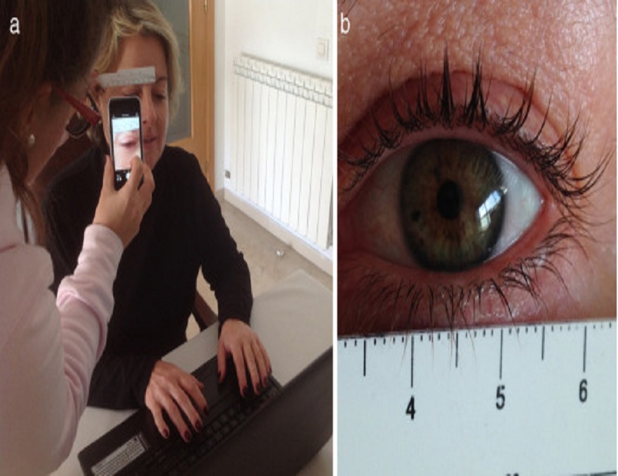
Pupil evaluation has always been integral to neuro exams, offering a unique, non-invasive window into the human brain. Recent advancements have rendered these assessments even more precise and insightful. Techniques for accurate pupil diameter measurement have evolved, turning pupillometry from a qualitative art into a quantitative science.
This article aims to provide medical professionals with an understanding of these advancements, shedding light on the fascinating journey from simple light reflex tests to the complex pupillary response analyses we have today.
Understanding Pupil Dilation and Constriction
One must first comprehend the basic pupil dilation and constriction processes to understand the nuances of pupillary evaluation. The autonomic nervous system manages these dynamic changes in pupil size, with dilation controlled by the sympathetic pathway and constriction by the parasympathetic pathway.
But beyond mere responses to light and darkness, pupillary changes also reflect diverse internal states and conditions. Everything from emotional arousal and cognitive load to systemic medications and the state of alertness can influence pupil size and reactivity. Recognizing these influences is fundamental in a neurological exam, distinguishing between physiological variation and potential signs of neurological compromise.
Advancements in Pupillometry
Pupillometry has come a long way from its traditional methods, thanks to advancements in technology and research. The manual observation of pupillary responses has given way to sophisticated devices capable of providing quantitative assessments. These devices can precisely measure various parameters, including pupil size, shape, symmetry, and reactivity, all of which have crucial roles in neurological assessment.
One such advancement is the Neurological Pupil Index (NPi), an objective, automated, and standardized measure of pupillary reactivity. Using a scale from 0 to 5, NPi enables clinicians to identify subtle changes in pupillary response that may precede clinical deterioration. Such early detection can be instrumental in managing neurocritical patients, positively impacting their prognosis and recovery.
Dynamic Pupillary Light Reflex Testing
Pupillary light reflex testing is a cornerstone of pupillary evaluation. The reflex involves a balance between sympathetic and parasympathetic activities and provides insights into the afferent and efferent pathways of the optic nerve and oculomotor nerve, respectively. However, traditional flashlight testing may not be sufficiently sensitive to detect subtle or early changes.
Dynamic pupillary light reflex testing solves this problem by evaluating pupillary responses over time and under varying light conditions. By providing a detailed analysis of pupillary constriction and dilation patterns, this technique can improve the sensitivity and specificity of neuro exams, aiding in the early detection and management of neurological conditions.
Assessing Relative Afferent Pupillary Defect (RAPD)
Detecting a Relative Afferent Pupillary Defect (RAPD) is integral to any comprehensive neuro exam. RAPD signifies an asymmetric functioning of the optic nerves, indicating potential conditions such as optic neuritis, severe retinal disease, or ischemic optic neuropathy.
Digital pupillometry has bolstered traditional methods like the swinging flashlight test, enhancing these evaluations’ accuracy, sensitivity, and specificity. The quantitative data obtained from these advanced techniques allow for more precise assessments, thereby improving the clinician’s ability to detect and monitor the progression of these conditions over time.
Quantitative Pupillography
Quantitative pupillography offers a more sophisticated analysis of pupillary behavior. This technique quantifies several pupillary light reflex parameters, including latency, amplitude, speed, and duration. It can dissect the pupillary response into distinct phases—like dilation and constriction—and provide a detailed analysis of each segment.
This level of detail allows for a comprehensive evaluation, uncovering potential abnormalities that may not be evident in traditional assessments. Quantitative pupillography can significantly enhance the diagnostic process with other neurological tools, providing invaluable insights into neurological status.
Conclusion:
The art of pupil evaluation has witnessed a significant evolution. With an array of advanced techniques and tools at our disposal, the accuracy and efficiency of neuro exams have dramatically improved. However, the heart of pupillary evaluation still lies in a fundamental understanding of pupillary behavior and its various influencing factors.
As medical professionals, we must stay abreast of these advancements and incorporate them into our practice. Furthermore, the potential of pupil evaluation in neuro exams is vast and unexplored, opening doors for future research and collaboration. As we step into this promising future, our commitment to improving the precision and quality of neuro exams must remain unwavering.

